As the body’s largest organ, the skin’s principal and most important function is as a barrier that prevents foreign substances and external stimuli from entering the body while regulating the loss of moisture from the body through evaporation. The deterioration of barrier function is known to lead to problems, including dry skin. According to a consumer survey, approximately 90% of women who reported the start of skin problems at around age forty, such as dryness associated with age, responded that their skin remains dry despite using skin care products.*1 In addition to providing moisturizing care for dry skin, Lion is developing technology to address skin concerns that enhance its natural strengths.
*1Survey of skin care awareness (November 2022, Lion survey of women aged 35–49, n = 2,283)
The skin is composed of a number of layers, including the epidermis, dermis, and hypodermal tissue, and the epidermis itself is composed of a horny layer, stratum granulosum, stratum spinosum and stratum basale (see Figure 1a). The horny layer is the outermost layer of the skin and acts as the first barrier, protecting the body from such external stimuli as ultraviolet rays and dryness. The stratum granulosum, located beneath the horny layer, contains “tight junctions.” Tight junctions (TJs) act as a second barrier and is involved in retaining water levels in the body (see Figure 1b).*2
*2Japanese Society of Aesthetic Dermatology. Modern Aesthetic Dermatology. Nanzando, 2022.
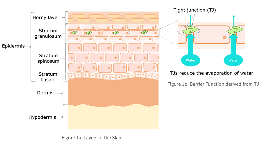
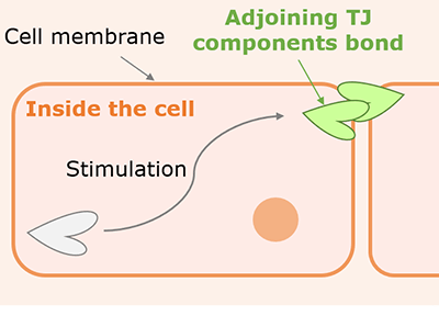
TJs are formed by multiple TJ component proteins, called TJ components, which are involved in cell adhesion. TJ components are produced inside of the cell and then transferred to the cell membrane. Adjacent TJ components bind to each other (shown in Figure 2), forming inter-cell adhesions and acting as a barrier to inhibit the passage of substances between cells (see Figure 1b).*3,4,5 To summarize, it is thought that TJ components on the cell membrane are an important component of the second barrier function. Therefore, our researchers looked for substances that can help increase the TJ components localized on the cell surface.
*3Kubo, A. Detailed understanding of the skin’s tight junction barrier structure. The Japanese Journal of Dermatology. Vol. 124, 3, 305–314, 2014.
*4French, A. D. et al. PKC and PKA phosphorylation affect the subcellular localization of claudin-1 in melanoma cells. Int. J. Med. Sci, 6, 93–101, 2009.
*5Kobayashi, M. et al. Weak ultraviolet B enhances the mislocalization of Claudin-1 mediated by nitric oxide and peroxynitrite production in human keratinocyte-derived HaCaT cells. Int. J. Mol. Sci, 21(19), 2020.
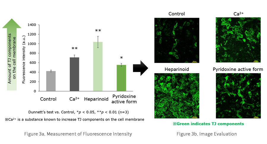
To identify what substances help increase TJ components in the cell membrane, Lion researchers used a novel high‑throughput screening system termed live-cell immunostaining. This is simple, highly accurate method of selectively staining the TJ components localized on the cell surface takes advantage of the cell’s adhesive properties and uses fluorescently labeled antibodies of the specific cell surface‑localized TJ components. One substance known to increase TJ components is Ca2+, and researchers added this and a number of potential candidate substances to epidermal keratinocytes, and then stained the TJ components through live-cell immunostaining. The results showed that fluorescence intensity increased significantly in heparinoid and vitamin B6 (pyridoxine active form) compared to the control group (see Figure 3a). Then, researchers evaluated the images to confirm that the green line (see Figure 3b) conformed to the shape of the cell membrane. These results indicated that there are two substances that can help increase the number of cell surface‑localized TJ components.
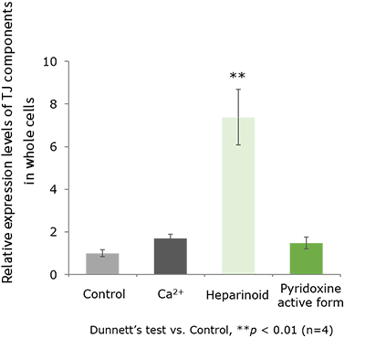
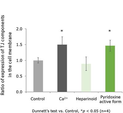
Next, to clarify the mechanism by which these substances help increase TJ components, the research team used the western blot technique to evaluate the expression of TJ components in the whole cell and proportion of TJ components in the cell membrane. First, they assessed the expression of TJ components in the whole cell, and their results showed a significant increase in the expression of TJ components when heparinoid was added. They also confirmed that the pyridoxine active form achieved expression equivalent to that of Ca2+ (see Figure 4). Next, they calculated the ratio of TJ component expression in the cell membrane compared to the whole cell and evaluated this ratio in comparison to the control ratio, which was set at 1. The results confirmed that the proportion of TJ components in the cell membrane significantly increased with the addition of the pyridoxine active form (see Figure 5). These results indicate that the pyridoxine active form is very effective in transferring TJ components into the cell membranes.
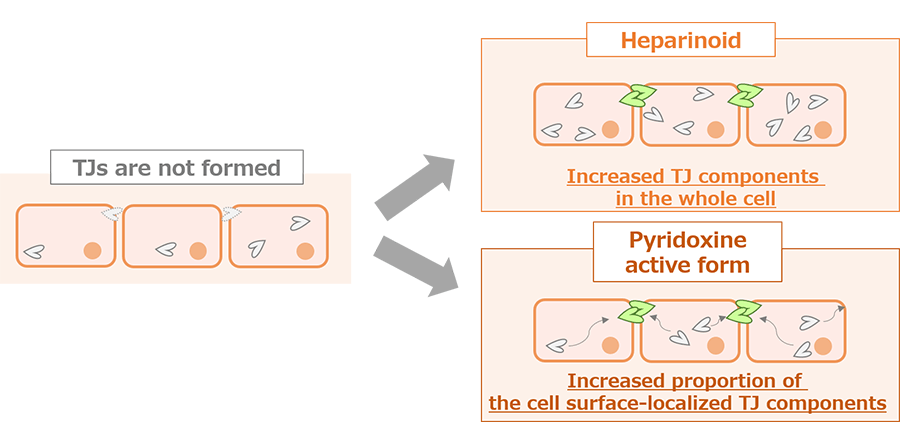
These findings suggest that heparinoid helps increase in TJ components throughout the cell and that the pyridoxine active form may specifically contribute to increasing the cell surface‑localized TJ components (see Figure 6).
At Lion, we believe that the key to treating dry skin is to go beyond providing moisture and to also draw out the skin’s natural ability to retain that moisture. We will continue to develop technologies that focus on the skin’s natural strength, such as research into TJs, to support better skin care habits for people who want to continue to look their best as they age.
Research Publication
Sakamoto, H. Nishikawa, M. et al. Development of tightjunction‑strengthening compounds using a high‑throughput screening system to evaluate cell surface‑localized claudin‑1 in keratinocytes. Sci. Rep. 14, 3312. (2024)License information for this article
These all data used under CC BY 4.0 / Cropped from originalHiroki Sakamoto, Momoyo Nishikawa, The 123th Annual Meeting of the Japanese Dermatological Association

Developing Pre-Caries Detection Technologies

Gum Disease Prevention Technology Development Focused on the Periodontal Tissue
Skin Research that Draws Out Skin’s Natural Strength

Study of a Specific Example of Genetic Polymorphism—The Relationship between the Function of Enteric-coated Lactoferrin and Single Nucleotide Polymorphisms (SNPs)

Genetic polymorphisms research to provide individualized healthcare based on each person's constitution
Related Information